

+918292345710

Currently it only shows your basic business info. Start adding relevant business details such as description, images and products or services to gain your customers attention by using Boost 360 android app / iOS App / web portal.
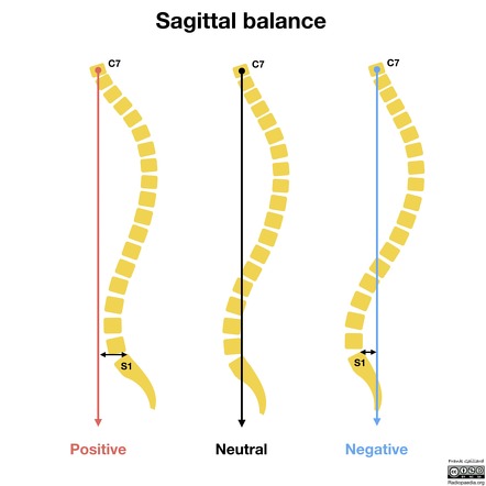
Radiological assessment of sagittal balance of spine by plain radiographs include upright anteroposterior and lateral full-length scoliotic views, long 17- by 36-inch cassette of the entire spine taken with the shoulders at 45-degree forward flexion and the hips and knees fully extended Full extension of the hips and knees are important to eliminate any compensatory flexion that may mask a severe deformity. Currently, the most used radiographic method to assess sagittal balance is the standing full-length lateral radiograph measuring the horizontal distance between a C7 plumb line and the posterosuperior aspect of the sacrum at the L5–S1 disk space, also known as sagittal vertical axis. Positive sagittal imbalance is defined as an anterior deviation of the C7 plumb line.
